Oxygen measurement in cells is the focus of science
October 9, 2019PreSens congratulates this year's Nobel Laureates and is pleased that attention has been paid to hypoxia in research in this way.
Cell oxygenation is at the heart of this year's Nobel Laureates research, scientists William Kealin Jr. (USA), Sir Peter Ratcliffe (UK) and Gregg Semenza (USA), who have discovered the molecular mechanisms by which cells measure and control the oxygen content to adjust.
An important role is played by the so-called hypoxia-induced factor (HIF), a transcription factor that regulates the supply of oxygen to the cells by balancing oxygen demand and oxygen supply. Among other processes, HIF significantly regulates erythropoiesis, angiogenesis and glucose transport. This "HIF switch" is one of the most important physiological adaptation reactions of the human body and shows that the oxygen environment should be considered in all cell research.
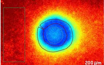
One of the big challenges of 3D fabric constructs for e.g. transplantation is the development of oxygen and nutrient gradients in the middle of the cultured tissue. The usual standard method in 3D cell culture, after cell cultivation, involves histological cutting and staining of the cells to detect oxygen distribution, hypoxia or even cell death. With its microsensors and the VisiSens™ Imaging System, PreSens offers the only measurement methods available to determine hypoxia online. They allow accurate O2 spot measurements in 3D cell constructs without self-consumption of oxygen and thus also draw conclusions on the oxygen-dependent regulation of HIF-dependent genes. Colgan et al. successfully demonstrate with PreSens measuring systems that the local HIF-mediated reduction of oxygen by migrating neutrophils positively influences the inflammatory response in intestinal inflammation [1].
The cell metabolism crucially depends on local O2 supply. Especially in 2D and 3D cell cultures or cultured tissue, cells that are located in diffusion-restricted regions (e.g. in cell carriers or spheroids) may be exposed to low oxygen concentrations. Non-invasive, continuous 2D mapping can be performed directly in the incubator under growth conditions. In addition, the distributions of analytes in living samples can be visualized in 2D.
The optical sensors from PreSens are already in use for the oxygenation monitoring of perfusates in various transplantation research projects.
Other potential applications include tumor oxygen monitoring / angiogenesis as well as the monitoring of vital organs, muscle tissue and heart valves, and oxygen measurements in ophthalmology.