
Watch tutorials, webinars and informative videos about PreSens optical sensor systems.

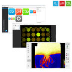
Oxygen Sensor Foil for Imaging Sample Surfaces or Through Transparent Vessel Walls
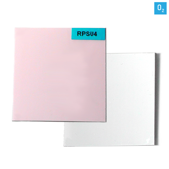
Oxygen Sensor Foil SF-RPSu4
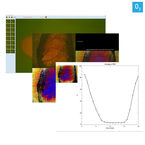
The SF-RPSu4 for measuring oxygen allows non-invasive mapping of metabolic activities as well as changes over time periods from seconds to months. The fluorescent sensor foil is attached on a living or dead sample surface or a transparent glass or disposable vessel. A sensor film on the foil translates the oxygen content into a light signal. The sensor foil is
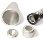
available in different sizes and can easily be cut in any desired shape. Read-out is done contactless with the imaging Detector Unit DU01 or the VisiSens TD.
- 2D read-out
- Contactless, direct sensing or through transparent walls
- Visualize spatial and temporal gradients
- Numerous measurement points in one image
Applications
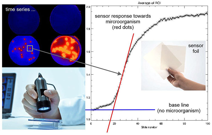
Detection of Microorganisms on Surfaces via O2 Imaging
Microorganisms which are responsible for nosocomial infections are known to be aeorbs or facultative anaerobs. This means that they consume oxygen due to their energy metabolism processes. In recent scientific studies, the VisiSens system has proofed to be able to monitor the very low oxygen consumption of microorganisms in 2D by sealing them between abiotical surfaces (e.g. desk) and the sensor foil. As a result, the presence of microorganism could be monitored in place within minutes.

O2, pH and CO2 Mapping in Sediments
O2, pH, and CO2 are key factors for microbial activity and various geochemical processes in sediments. They highly vary locally, e.g. at interfaces or different depths. Spatial and temporal analyte dynamics over long time periods can be visualized. Various regions can be compared within one measurement. VisiSens enables non-invasive 2D-mapping over cross-sections or on sample surfaces. The portable device can be used in lab and field.
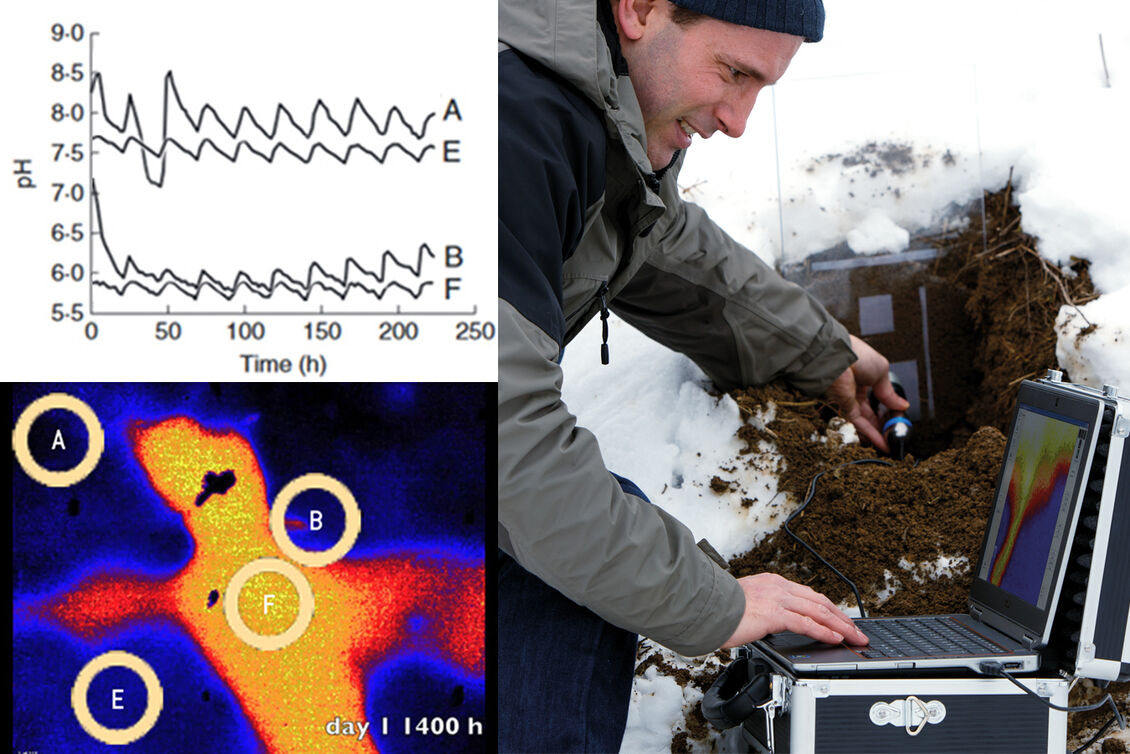
Spatial and Temporal Analyte Changes in Plants & Soil
O2, pH and CO2 play a crucial role in plant and soil processes, e. g. in photosynthesis, respiration, in rhizospheres or in microbiological processes. Metabolic processes can be monitored. This planar optical sensor technique allows non-invasive read-out through glass walls of rhizotrons. Studying metabolic activity of roots and determining the cultivation optimum is important for sustainable agriculture, e. g. for adjustment of water and fertilizer supply.
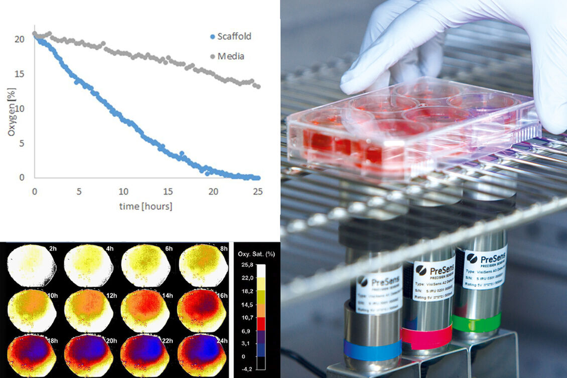
O2 or pH in Cell Culture and Engineered Tissue
Cellular metabolism critically depends on local O2 supply and pH values. Especially in 2D and 3D cell culture or engineered tissue, cells located in diffusion limited regions (e.g. in scaffolds or spheroids) can be subject to low oxygen levels and pH changes. Non-invasive, continuous 2D-mapping can be performed directly in the incubator under growth conditions. Furthermore, 2D analyte distributions in living samples can be visualized.
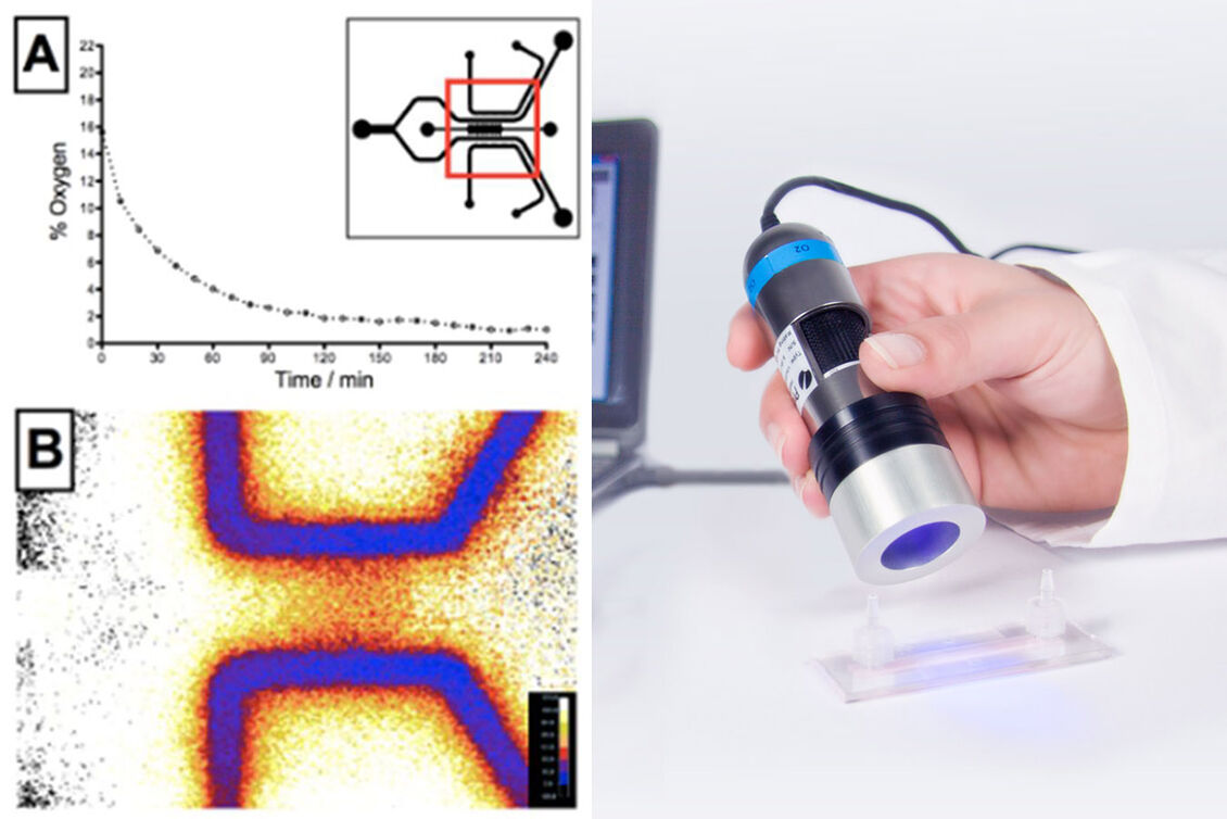
Non-invasive 2D Analyte Mapping in Microfluidics
VisiSens™ enables 2D visualization of important culture parameters inside microfluidic chips. You can continuously monitor in 2D, with high resolution at specific positions or over the whole chip surface in a non-contact readout mode. Detect metabolic hotspots, record time-series, and monitor hypoxia, cellular growth, or O2 supply inside the chip. You can gain new insights on metabolic activity and natural or artificially produced gradients.
Technical
| Specifications# | |
|---|---|
| *typical data which may strongly differ with adapting the imaging set-up to specific needs **typical data of LOD of a defined ROI (> 6,000 pixels) over time at + 20 °C, excluded ambient light, FoV 8 cm x 6 cm, VisiSens DU01 strongly differs ***typical data of precision of a defined ROI (> 6,000 pixels) over time at + 20 °C, excluded ambient light, FoV 8 cm x 6 cm, VisiSens DU01 strongly differs **** typical data of spatial standard deviation in defined ROI (> 6,000 pixels) at + 20 °C, excluded ambient light, FoV 8 cm x 6 cm, VisiSens DU01 strongly differs #VisiSens™ is no approved medical device | |
| Measurement range | 0 – 100 % air saturation(0 – 20.9 % O2) |
| Response time* (t90) | Gas phase: < 8 sec. Dissolved: < 30 sec. |
| Specifications using VisiSens TD read-out | |
| Limit of detection** | 0.03 % air saturation |
| Precision (temporal)*** | ± 0.02 % air saturation at 0 % air saturation ± 0.1 % air saturation at 100 % air saturation |
| Precision (spatial)**** | ± 1.5 % air saturation at 0 % air saturation ± 3.0 % air saturation at 100 % air saturation |
| Properties | |
| General sensor temperature working range | from + 5 to + 45 °C |
| Compatibility | Aqueous solutions, ethanol (max. 10 % v/v), methanol (max. 10 % v/v), pH 2 - 10 |
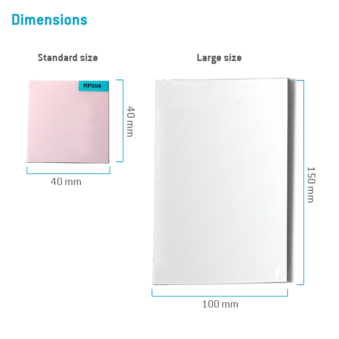
| Size of sensor foil | Standard 40 x 40 mm² Min. 5 x 5 mm² |
Related products
Resources
Publications
O2-sensitive Microcavity Arrays for 3D Cell Culture Monitoring
In situ Remediation of Contaminated Groundwater
Monitoring Spatio-Temporal Oxygen Consumption Within Live 3D Cancer Spheroids
How Much O2 Do Cells Really Face?
Spatio-temporal O2 Gradients in the Microenvironment of an Outgrowing Cell Patch
Oxygen Consumption Rates of Adherent MDCK II Monolayer Cells
Monitoring Oxygenation in Microfluidic Cell Culture Using 2D Sensor Foils as Growth Substrate (full text)
Adhesive Cell Growth and Microscopy on O2 and pH Sensor Foils
Imaging of pH and pO2 on Irradiated Fibroblasts and Oral Squamous Carcinoma Cells
Cellular Oxygen Consumption in Microfluidic Devices
Role of Oak Wood Ellagitannins on the Oxygen Transfer Rate in Wine Barrels
Benthic Disturbance-Recovery Dynamics in a Changing Coastal Ocean
Immobilized Plant Cells - From DSMZ to the Customer
Diffusive / Dispersive Reaction Fronts in Groundwater
Exchange of O2 and CO2 in the Capillary Fringe of a Porous Medium
Monitoring Oxygen in a Mouse Model of Renovascular Hypertension
Measuring Respiration and Primary Production Dynamics in Macroalgae
Assessment of Oxygen Depletion and Biofilm Structure Grown in MBBR Carriers
Measuring Oxygen Gradients above a Respiratory Active Algal Cell Layer
Viability of Lithobiontic Microorganisms Inhabiting the Rocks of Atacama Desert
Oxygen Dynamics in the Capillary Fringe
Monitoring Skin Tissue Oxygenation in Mice
Recording Spatial Patterns of Oxygen Consumption in Individual Corals
Effect of Non-Symbiotic Hemoglobin Expression on Oxygen Content in Roots
Effect of Pseudomonas Infection on Oxygen Status of Arabidopsis Leaves
Integration of Oxygen Sensor Foils in Microfluidic Chips
O2, CO2 and pH Dynamics in the Capillary Fringe
Imaging the Oxygen Consumption of Microbial Cultures
Monitoring pO2 in Cell Culture to Improve Directed Differentiation
Oxygen Dynamics around Buried Tar Balls in Florida Marine Sands
Assessing Biocide Actions on Lichen with VisiSens
Perfusion Monitoring in Microvascular Flaps
Extracorporeal Membrane Oxygenator to Support Diseased Lungs
Quantification of oxygen consumption in head and neck cancer using fluorescent sensor foil technology
O2-sensitive microcavity arrays: A new platform for oxygen measurements in 3D cell cultures
Physiological oxygen measurements in vitro – Schrödinger´s cat in 3D cell biology
O2-sensitive Mikrokavitätenarrays: 3D-Zellkultursystem mit Sensorfunktion
Plant-Mediated Rhizosphere Oxygenation in the Native Invasive Salt Marsh Grass Elymus athericus
A New Non-invasive Technique for Measuring 3D-Oxygen Gradients in Wells During Mammalian Cell Culture
Mapping the biological activities of filamentous oxygenic photogranules
In situ observation of pH change during water splitting in neutral pH conditions: impact of natural convection driven by buoyancy effects
A Microfluidic Chip Architecture Enabling a Hypoxic Microenvironment and Nitric Oxide Delivery in Cell Culture
Impact of Bed Form Celerity on Oxygen Dynamics in the Hyporheic Zone
The effect of unsteady streamflow and stream-groundwater interactions on oxygen consumption in a sandy streambed
A novel approach to investigate hypoxic microenvironment during rice colonization by Magnaporthe oryzae
Microfluidic Patterned Model of Non-alcoholic Fatty Liver Disease: Application to Disease Progression and Zonation
Scalable Microfluidic Platform for Flexible Configuration of and Experiments with Microtissue Multiorgan Models
Iron Lung: How Rice Roots Induce Iron Redox Changes in the Rhizosphere and Create Niches for Microaerophilic Fe(II)-Oxidizing Bacteria
More than Meets the Dye: Evaluating Preferential Flow Paths as Microbial Hotspots
Elevated micro-topography boosts growth rates in Salicornia procumbens by amplifying a tidally-driven oxygen pump: implications for natural recruitment and restoration
Imaging of pH and pO2 gives insight in molecular processes of irradiated cells
Experimental study on the oxygenation efficiency of nano-bubble modified mineral particles at the sediment-water interface in lakes
Successful control of phosphorus release from sediments using oxygen nano-bubble-modified minerals
Hypoxic Physiological Environments in a Gas-Regulated Microfluidic Device
Redox regulation of EGFR steers migration of hypoxic mammary cells towards oxygen
Dynamics of oxygen and carbon dioxide in rhizospheres of Lobelia dortmanna - a planar optode study of belowground gas exchange between plants and sediment
Oxygen transport in periodically ventilated polychaete burrows
2D visualization captures the local heterogeneity of oxidative metabolism across soils from diverse land-use
High Resolution Assessment of Spatio-Temporal Changes in O2 Concentration in Root-Pathogen Interaction
Oxygen-distribution within 3-D collagen I hydrogels for bone tissue engineering
A microfluidic oxygen sink to create a targeted cellular hypoxic microenvironment under ambient atmospheric conditions
Plant-Sediment Interactions in Salt Marshes - An Optode Imaging Study of O2, pH, and CO2 Gradients in the Rhizosphere
Spatial organization of bacterial populations in response to oxygen and carbon counter-gradients in pore networks
Linking biofilm spatial structure to real-time microscopic oxygen decay imaging
Anoxic microsites in upland soils dominantly controlled by clay content
An experimental-numerical investigation on the effects of macroporous scaffold geometry on cell culture parameters
Who really matters: Influence of German Bight key bioturbators on biogeochemical cycling and sediment turnover
Oxygen Mapping: Probing a Novel Seeding Strategy for Bone Tissue Engineering
Wurzelexsudation in der Wasserfluktuationszone des Drei-Schluchten-Reservoirs, VR China. Ein Ansatz für Einblicke in eine überflutungsgestresste Rhizosphäre am Beispiel von Salix variegata Franch
Effects of light fractionation and different fluence rates on photodynamic therapy with 5-aminolaevulinic acid in vivo
Transcutaneous pO2 imaging during tourniquet-induced forearm ischemia using planar optical oxygen sensors
Determination of Oxygen Gradients in Engineered Tissue Using a Fluorescent Sensor
Multi-ion imaging using fluorescent sensors in a microtiterplate array format
Fluorescence Detection and Diagnosis of Non-Melanoma Skin Cancer at an Early Stage
Fluorescent Imaging of pH with Optical Sensors Using Time Domain Dual Lifetime Referencing
Luminescence Lifetime Imaging of Oxygen, pH, and Carbon Dioxide Distribution Using Optical Sensors
Luminescence Lifetime Temperature Sensing Based on Sol-Gels and Poly(acrylonitrile)s Dyed with Ruthenium Metal-Ligand Complexes
Simultaneous Imaging of Cortical Partial Oxygen Pressure and Anatomic Structures Using a Transparent Optical Sensor Foil
Transcutaneous pO2 measurement during tourniquet-induced venous occlusion using dynamic phosphorescence imaging
A new semi-invasive method for two dimensional pO2 measurements of cortical structures
Non-invasive measurement of the superficial cortical oxygen partial pressure
Radial oxygen gradients over rat cortex arterioles
Planar Oxygen Sensors for Non Invasive Imaging in Experimental Biology
Visualisation of Cortical pO2 During an Epidural Mass Lesion in Rodents
Simple, fast and reliable perfusion monitoring of microvascular flaps
An imaging method for oxygen distribution, respiration and photosynthesis at a microscopic level of resolution
Characterization of a chip-based bioreactor for three-dimensional cell cultivation via magnetic resonance imaging
Ratiometric luminescence 2D in vivo imaging and monitoring of mouse skin oxygenation
Real-Time 2D Visualization of Metabolic Activities in Zebrafish Embryos Using a Microfluidic Technology
Low-oxygen tensions found in Salmonella-infected gut tissue boost Salmonella replication in macrophages by impairing antimicrobial activity and augmenting Salmonella virulence
Online Monitoring of Crude Oil Biodegradation at Elevated Pressures
Effect of Farnesyltransferase Inhibitor R115777 on Mitochondria of Plasmodium falciparum
Ultrasonic welding of chemical optical sensors supporting O2, pH and CO2 imaging in microfluidic systems
Oxygen measurement in interstitially perfused cellularized constructs cultured in a miniaturized bioreactor
FAQs
Is it necessary to coat VisiSens sensor foils with cell adhesion promoting proteins, and how do I coat them?
My live image representation in the VisiSens software is very slow, and I dont´t get a smooth reproduction. What can I do?
The standard size of the VisiSens sensor foils is rectangle 4 x 4 cm. What can I do, if my sample requires a different size or shape?
There are "jumps" in my VisiSens time series. What can I do?
What is the maximum resolution of VisiSens A1? Can we see up to micro-scale?
What is the minimum sensor size for VisiSens?
What is the time of delivery?
Which side of the VisiSens sensor foils has to face the sample?
Which substances can interfere with the optical O2, pH and CO2 measurements?
Manuals
O2 Sensor Foil SF-RPSu4
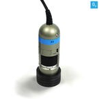
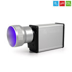
Detector Units DU01 / DU02 / DU03
VisiSens AnalytiCal 1
Media
Video: VisiSens - Oxygen Distributions in E. coli Culture
Video: VisiSens - Visualizing Biocide Actions on Lichen
Video: VisiSens - Oxygen Imaging in Microfluidics
Video: VA1 Basic Functions
Video: VA1 Measurement
Video: VA1 Calibration
Video: VisiSens Delivered Equipment
Video: How2 Measure and Visualize O2, pH and CO2 in 2D for Biological Research
Video: How2 Measure and Visualize O2, pH and CO2 in 2D for Life Science Research
Video: VisiSens WEBINAR - Metabolic Activity inside Microfluidics
Video: VisiSens WEBINAR - O2, pH & CO2 in Sediments, Interfaces and Biofilms
Video: VisiSens WEBINAR - O2, pH & CO2 in Plants, Roots and Soil
Video: VisiSens WEBINAR - O2 & pH in Cell Culture, Engineered & Native Tissue
Media Release: Oxygen in Action
Media Release: Microfluidic Devices