

Watch tutorials, webinars and informative videos about PreSens optical sensor systems.
Measuring Respiration and Primary Production Dynamics in Macroalgae
2D imaging of oxygen gradients in F. vesiculosus with the VisiSens system

K. Spilling
Finnish Environment Institute, Marine Research Laboratory, Helsinki, Finland
In this study the VisiSens system was used for 2D imaging of small scale differences in primary production and respiration in macroalgae. F. vesiculosus was used as a model organism. With the VisiSens system a clear difference in respiration and primary production between pieces from different parts of the thallus could be detected. Respiration rate and production were lowest near the holdfast and increasing towards the apical tips where metabolic activity was highest. Even small scale respiration `hotspots´ of 100 μm in diameter could be detected at the apical tips. Compared to selective measurements with Clark-type microelectrodes, which had been used in previous sutdies, spatial differences are easier to detect and oxygen gradients can be examined quicker and with greater accuracy by recording 2D oxygen images with the VisiSens system.
There is hardly any information about the interplay between the structure of algal tissue and its effect on physiological activity. Recently we showed that there can be considerable differences in photosynthetic activity and respiration on a microscopic spatial scale in seaweed. However, very few studies of spatial differences in physiological activity in macroalgae on micro-scale exist. Previous investigations of oxygen micro-gradients in macroalgae have relied on Clark-type oxygen microsensors involving very time consuming measurements. The benefit of using a 2D system such as VisiSens is to create images where spatial differences are easier to detect and where oxygen gradients can be examined quicker and with greater accuracy compared to several pinpoint measurements. In this study the suitability of the VisiSens system for detecting small-scale differences in respiration and primary production in macroalgae was tested, and the common bladder wreck, Fucus vesiculosus, was used as a model species. F. vesiculosus is a perennial macroalgae that has several distinct parts such as holdfast, mid stem and apical parts (see Fig. 1). All different parts and thereby different tissue structures have been investigated with VisiSens.
Materials & Methods
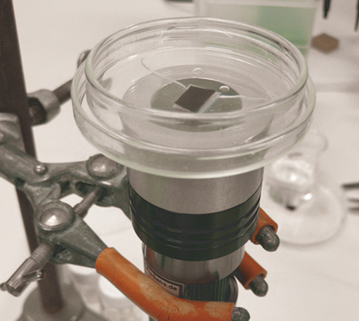
Specimens of F. vesiculosus (bladder wreck) were taken from shallow depth (0.5 m) on the southern side of the Lautasaari island, Helsinki. The algae were taken to the lab and gradually acclimated to warmer temperature in a water bath. After acclimation to room temperature (22 °C), the algae were stored in a container with natural sea water aerated with air in order to keep the oxygen concentration close to 100 %. The algae were placed in light of about 20 µmol photons m-2 s-1, in a 16 h : 8 h light-dark cycle. Evaporation water from the storage container was replaced every day with addition of Milli Q water. Small pieces of F. vesiculosus thallus, from different parts of the seaweed, were cut out and placed on top of the oxygen sensor foil (SF-RPSU4, PreSens) inside a glass Petri dish. The lid part of the Petri dish was used for this purpose and the `box´ part was placed on top to keep downward pressure on the thallus. This was done in order to prevent air bubbles between the thallus and the sensor foil, and to have the thallus evenly pressed towards the sensor foil. The VisiSens camera system was placed facing upwards using a lab arm and the petri dish was placed on top (see Fig. 2). The measurement cycle started by determining the oxygen concentration every 60 s in darkness. After 7 minutes in the dark the thallus was illuminated for 5 s with 800 µmol photons m-2 s-1 with a white LED. Oxygen was determined after each illumination period and followed by 5 s in the dark. After 17 cycles with 5 s illumination, the light period was increased to 30 s, and 7 cycles of longer light period creating a total of 30 images were recorded.
Respiration and Production Rate
There was a clear difference in respiration and primary production between pieces from different parts of the thallus. Respiration rate and production rate were lowest near the holdfast and increasing towards the apical tips where the metabolic activity was highest (Fig. 3 A + B, Fig. 4 A). At the apical tips there were also distinct differences on a small spatial scale, which had clearly higher respiration rate and lower production rate (Fig. 4 B). These small scale respiration `hotspots´ were in the order of 100 µm in diameter. These areas with higher respiration rate could be related to the cryptostomata structures that have been suggested to have elevated respiration rate, but it is clear form this investigation that not all the cryptostomata have this, as several apparently did not have any different metabolic activity from the surrounding thallus tissue (Fig. 4 A + B).


Conclusion
The VisiSens system provided images of oxygen concentrations at µm scale. There are measurable differences in metabolic activity between different parts of F. vesiculosus thallus. With 2D imaging it was possible to detect oxygen gradients, and relate areas with higher respiration rate to certain tissue structures. Compared to the time consuming measurements with Clark-type oxygen microsensors VisiSens enables easier detection and quicker examination of oxygen gradients with higher accuracy.