Watch tutorials, webinars and informative videos about PreSens optical sensor systems.
Integration of a PreSens Oxygen Sensor Spot Into a Solid Mini Bioreactor
Validation of a Newly Designed Sensor Adapter
M. Eng. Nico Wüstneck
Department Cell Techniques and Applied Stem Cell Biology, University Leipzig, Germany
This report illustrates the operation of a sensor adapter using PreSens sensor technology to measure environmental conditions inside a non-transparent solid mini bioreactor. With this approach the application remains easy to handle and different types of optical sensors (O2, pH, or CO2) can be applied or exchanged quickly on demand without modifying the bioreactor itself.
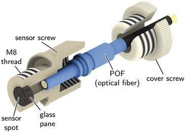
The Department Cell Techniques and Applied Stem Cell Biology of the University of Leipzig developed a mini bioreactor (diameter: 69 mm, height: 39 mm) to investigate and enhance the production of hyaline cartilage transplants in-vitro [2]. For this purpose it is necessary to control and measure selected parameters of the cell culture medium which surrounds the grafts inside the bioreactor. While, for example a decrease in pH value could clearly indicate an infection, a decrease in dissolved oxygen would show increased cell activity. Beside the relation to cell vitality the oxygen rate can be a fixed condition. In contrast to blood, where oxygen tension is around 20 %, it is only 1 - 6 % in the cartilage of the human knee [1]. To run trials with different predefined oxygen tensions there is a need to monitor this parameter. For several reasons the bioreactor is made of a non-transparent thermoplast. In order to use chemical optical sensors and read the sensor response non-invasively with the respective oxygen meter from the outside of the bioreactor an optical, transparent window would be needed. To realize this, a sensor adapter (Fig. 1) which can be attached to the bioreactor, was developed. To evaluate the bioreactor in combination with an oxygen sensor adapter, two experiments were performed. The first test demonstrated the dynamic behavior of the sensor, when an oxygen absorber was added to a fluid inside the bioreactor. The second experiment was conducted to simulate a very high cell activity, in order to validate the bioreactors capability to increase the oxygen tension of the fluid.
Materials & Methods
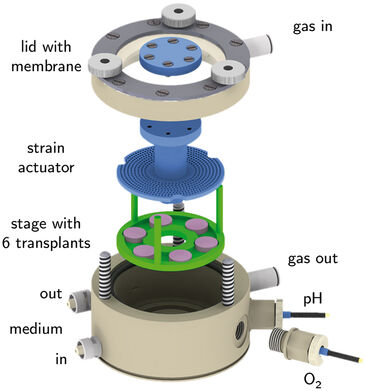
To simulate different conditions the bioreactor (Fig. 2) was designed to apply mechanical forces on the transplants, refresh the culture medium - which in addition applies shear forces - and control the oxygen level inside. The compression of the transplants - to stimulate cell activity - is done by a strain actuator with a discoidal plate, which is attached at the transparent and flexible silicon membrane of the lid. From above an external drive is magnetically docked to the actuator and manipulates its vertical position. One positive side effect of the mechanical stimulation is the swirl of the culture medium. As a consequence a well-balanced distribution of dissolved gas and nutrients inside the medium can be assumed. The lowest pair of in- and outlets on the side of the bioreactor is used for medium exchange, while the upper pair is intended for introduction of gas, which diffuses into the medium. Like most parts of the bioreactor the sensor adapter is made of the biocompatible thermoplast TECAPEEKTM. Equipped with a M8 thread it is laterally attached to the bioreactor level with the transplants. A glass pane (BOROFLOATTM; diameter: 5mm, height: 1.75 mm) is pasted to the front edge of the adapter with silicon. The used oxygen sensor spot (type SP-PSt3-YAU-D5-YOP, PreSens GmbH) is fixed on the glass pane with SG1-silicon glue. The polymer optical fiber (POF), which transfers excitation light to the sensor and the sensor response to the oxygen meter (Fibox 3, PreSens GmbH) can be inserted into the adapter from the opposite side of the glass pane for non-invasive measurements. A cover screw fixes the POF very closely to the glass pane.
Both experiments were performed with 17 mL of a common culture medium (DMEM) which is a composition of salts, vitamins, amino acids and glucose. The stage and transplants were not applied in the bioreactor for these tests. To compare DMEM with another fluid demineralised water was used. For the oxygenation experiment the introduced gas contained O2 (20 %), CO2 (5 %), and N2 (75 %) and removal of O2 from the fluids was done by adding 2 g of sodium sulfite (Na2SO3): 2 Na2SO3 + O2 -> 2 Na2SO4
Measurements in the Mini Bioreactor
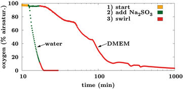
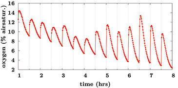
Fig. 3 shows the difference in oxygen decrease for both water and DMEM in the first experiment, if the same amount of Na2SO3 was added. After 5 min the strain actuator of the closed bioreactor was started to speed up the chemical reaction. Assuming an exponential decrease of oxygen the time constant for water (3 min) and DMEM (100 min) differs by a factor of more than 30. The reason for this could be a much slower solubility of Na2SO3 in DMEM because no changes could be detected till the swirl was caused by the strain actuator. Fig. 4 displays the cyclic oxygenation of Na2SO3 saturated DMEM with 20 % O2 for 3 min, which was performed with 200 mL/min every 30 min. The dissolution of Na2SO3 was still in progress so the O2 level tended to decrease. But there is also a clear indication for a quick impact of oxygenation. Summarized both experiments verified that the sensor adapter caused no constraints according to handling and performance of the bioreactor. The PreSens oxygen sensor spot proofed as fully functional with sufficient dynamic behavior in this set-up. Firstly an amplitude of the sensor signal between 30.000 and 70.000 ensures stable and precise measurement and secondly the adapter leaves the bioreactor as a closed and sterile system.
References:
[1] Gibson J. S., Milner P. I., White R., Fairfax T. P. A., Wilkins R. J.: Oxygen and reactive oxygen species in articular cartilage: modulators of ionic homeostasis. Pflüger Archiv - European Journal of Physiology 455 (2007), Nr. 4, 563 - 573
[2] Schulz R. M., Wüstneck N., van Donkelaar C. C., Shelton J. C., Bader A.: Development and validation of a novel bioreactor system for load- and perfusion-controlled tissue engineering of chondrocyte-constructs. Biotechnology and Bioengineering 101 (2008), Nr. 4, 714 - 728

