New Insights into Oxygen Supply in 3D Cell Cultures
December 12, 2023Knowledge about the oxygenation status of different zones within multicellular tumor spheroids is of essential interest for tumor research. The oxygenation status may serve as an indicator for the efficacy of anticancer drugs and can help to identify and choose suitable and personalized drugs.

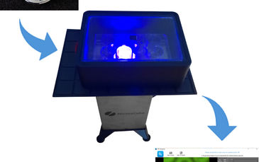
Prof. Dr. Wegener and his team from the University of Regensburg used a biocompatible measurement technique based on the VisiSens TD MIC System to measure O2 gradients in a cross section of a living spheroid. With a special microscopic imaging add-on they recorded 2D cross-sectional oxygen profiles of MCF-7 spheroids over longer periods of time.
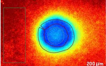
Therefore, a single 3D cultured spheroid is placed on the surface of an oxygen sensor foil. There it builds a semi-spherical structure (Fig. A) that enables recording of direct cross-sectional 2D oxygen maps from the contact zone between the spheroid and the sensor foil and visualizes any gradients within (Fig. B).
In contrast to usual measurement methods (e.g. staining with indicator dyes or nanoparticle sensors) the minimal-invasive oxygen mapping technique presented here can reach inner parts of the 3D cell cluster and maintains the structure and integrity of the spheroid.
Would you also like to monitor spatial and temporal oxygen distributions in 3D cell culture models under growth conditions? For more information read our application note on “Monitoring Spatio-Temporal Oxygen Consumption Within Live 3D Cancer Spheroids”.


