Watch tutorials, webinars and informative videos about PreSens optical sensor systems.
Detector unit for 2D pH imaging
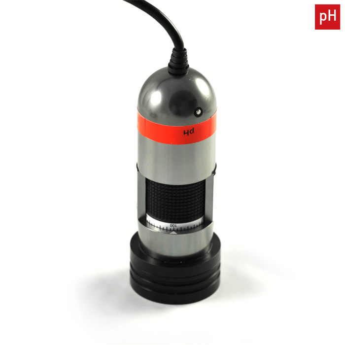
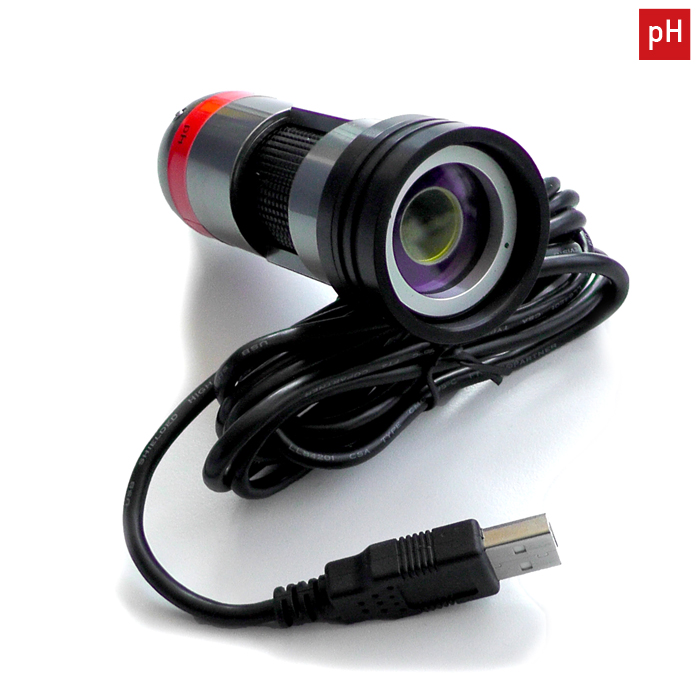
Detector Unit DU02
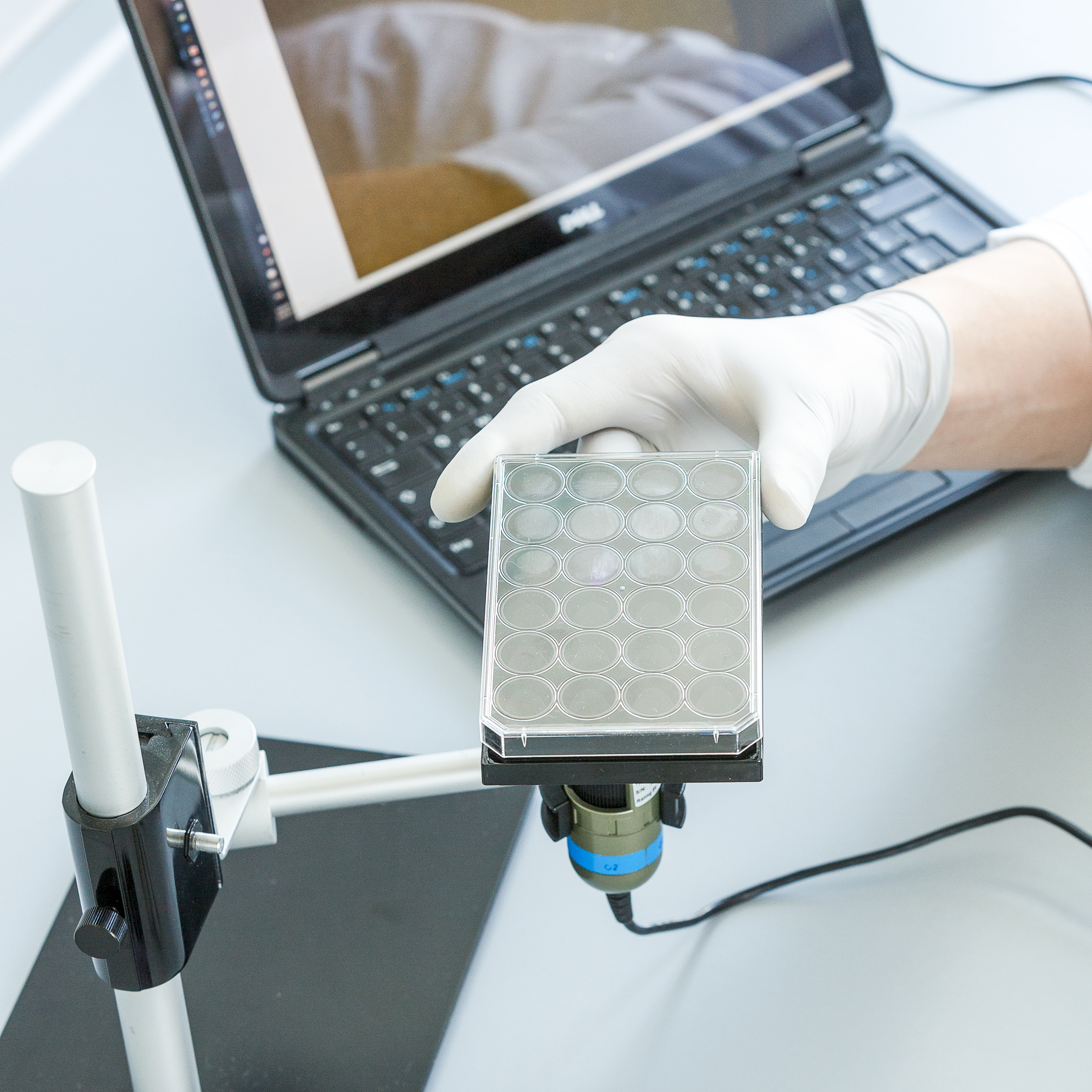
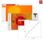
The VisiSens Detector Unit DU02 for pH imaging consists of an easy to use handheld digital camera, and a control and evaluation software. Combined with fluorescent chemical optical sensor foils, this imaging technology allows easy 2D visualization of pH distributions in heterogeneous samples. The software allows controlling the image recording process and assists image processing and evaluation. An easy to use camera controlling user interface manages image acquisition and storage. Measurements which belong together can be organized as user defined sessions in separate folders and annotated with a free text comment.
- Read-out of pH sensor foils
- More than 100,000 measurement points within one recorded image
- USB-powered portable microscope detector unit
- Small to medium size field of view (4.6 mm² to 13.5 cm²)
- Image processing and evaluation software included
- Visualize spatial and temporal gradients
- Time-lapse analyte movies
Applications
O2, pH and CO2 Mapping in Sediments
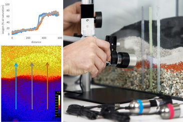
O2, pH, and CO2 are key factors for microbial activity and various geochemical processes in sediments. They highly vary locally, e.g. at interfaces or different depths. Spatial and temporal analyte dynamics over long time periods can be visualized. Various regions can be compared within one measurement. VisiSens enables non-invasive 2D-mapping over cross-sections or on sample surfaces. The portable device can be used in lab and field.
Spatial and Temporal Analyte Changes in Plants & Soil
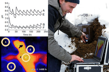
O2, pH and CO2 play a crucial role in plant and soil processes, e. g. in photosynthesis, respiration, in rhizospheres or in microbiological processes. Metabolic processes can be monitored. This planar optical sensor technique allows non-invasive read-out through glass walls of rhizotrons. Studying metabolic activity of roots and determining the cultivation optimum is important for sustainable agriculture, e. g. for adjustment of water and fertilizer supply.
O2 or pH in Cell Culture and Engineered Tissue
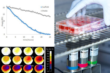
Cellular metabolism critically depends on local O2 supply and pH values. Especially in 2D and 3D cell culture or engineered tissue, cells located in diffusion limited regions (e.g. in scaffolds or spheroids) can be subject to low oxygen levels and pH changes. Non-invasive, continuous 2D-mapping can be performed directly in the incubator under growth conditions. Furthermore, 2D analyte distributions in living samples can be visualized.
Non-invasive 2D Analyte Mapping in Microfluidics
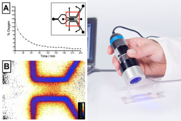
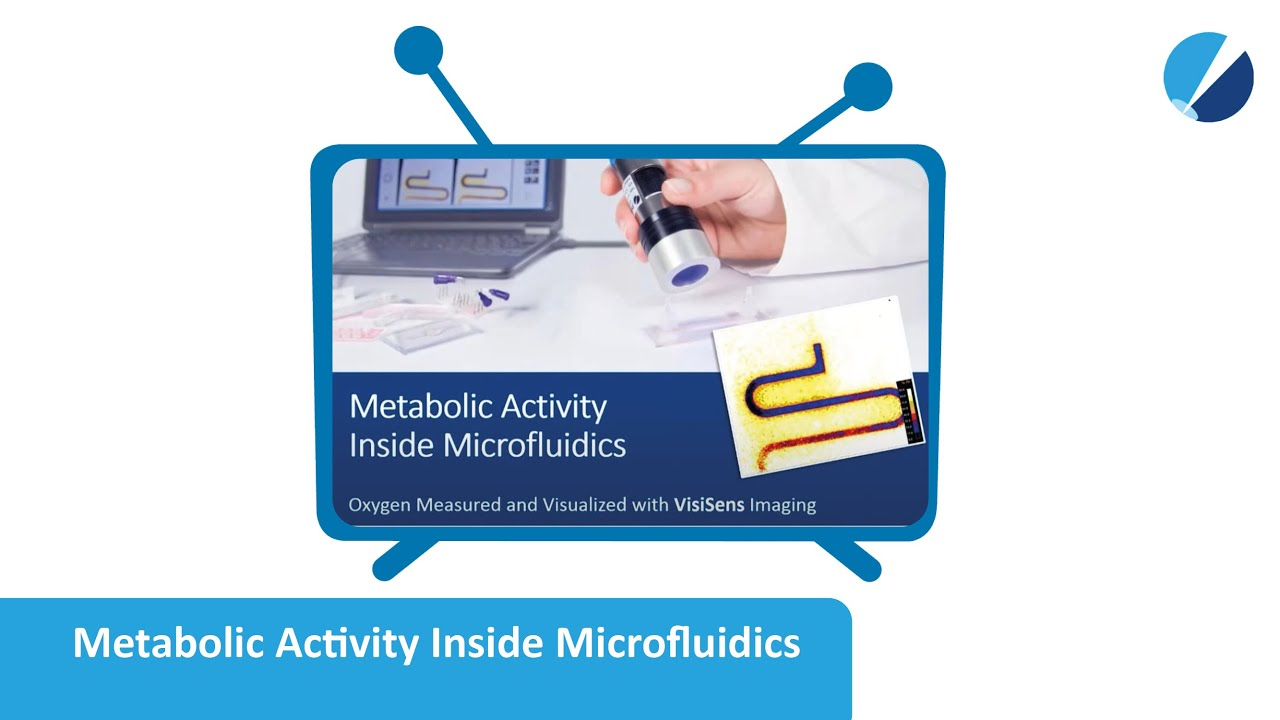
VisiSens™ enables 2D visualization of important culture parameters inside microfluidic chips. You can continuously monitor in 2D, with high resolution at specific positions or over the whole chip surface in a non-contact readout mode. Detect metabolic hotspots, record time-series, and monitor hypoxia, cellular growth, or O2 supply inside the chip. You can gain new insights on metabolic activity and natural or artificially produced gradients.
Technical
| Specifications | |
|---|---|
| Camera chip | Enhanced Color CMOS |
| Image resoltuion | 1.3 megapixel (1280 x 1024 pixels) |
| Magnification | 10-fold up to 220-fold, depending on adapter tubus used |
| Field of view | ∼ 1.6 x 1.3 mm2 to ∼ 3.6 x 3.0 cm2; typically ∼ 1.2 x 1.0 cm2 |
| Output | 15 fps live video preview (no storage) and 0.5 fps full-resolution picture storage (.png) |
| Interface | USB 2.0, high speed USB transmission |
| Number of LEDs | 8 |
| Material | All-aluminum housing |
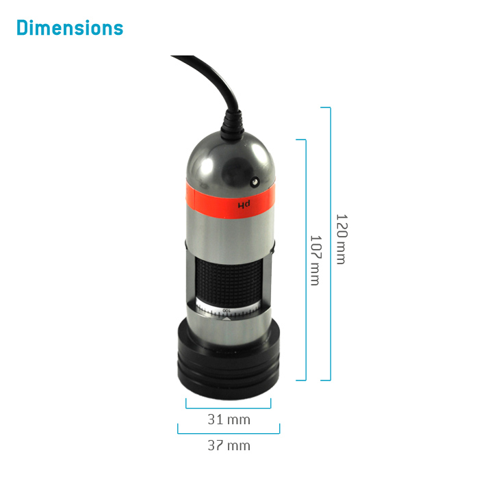
| Dimensions | Length 10 cm, diameter 3.8 cm |
| Weight | 170 g (without adapter tubus) |
Related products
Resources
Publications
FAQs
Media
Video: How2 Measure and Visualize O2, pH & CO2 Distributions in 2D for Biological Research
Video: How2 Measure and Visualize O2, pH & CO2 Distributions in 2D for Life Science Research
Video: VisiSens Delivered Equipment
Video: VisiSens WEBINAR - O2, pH & CO2 in Sediments, Interfaces and Biofilms
Video: VisiSens WEBINAR - O2, pH & CO2 in Plants, Roots and Soil
Video: VisiSens WEBINAR - O2 & pH in Cell Culture, Engineered & Native Tissue

