2D Oxygen Imaging in Micro-Scale with the VisiSens TD MIC System
September 28, 2020Now you can analyze oxygen distributions, gradients and hot spot formation in your sample 2-dimensionally, in fine scale resolution. The VisiSens TD MIC system can be adapted with optics and excitation light sources for microscopic oxygen imaging.
Our VisiSens TD MIC system is the ideal tool to study pO2 levels in 2D cell culture or other microscopic samples. Cell cultures are often grown under standard incubator conditions, although hypoxia is common to many types of tissue in the body. Therefore, it is important to monitor and analyze the oxygen levels in the direct microenvironment of the cells.
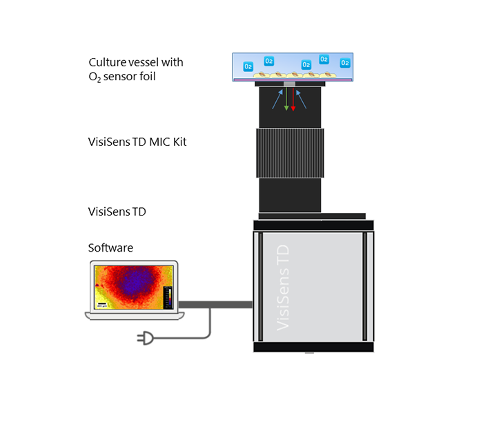
The VisiSens TD expanded with the MIC Kit fits into an incubator so you can study your samples under variable conditions. Core of the system is the oxygen-sensitive sensor foil, which you can place in your culture dish or on a glass slide. The sensor can be used as culture substrate for adherent cells. The camera component is put underneath the sensor and imaging can be performed without direct contact. Due to the proximity of cells and sensor you will be able to non-invasively measure temporal and spatial changes of O2 gradients caused by the actively respiring cells.
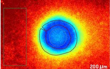
If you are interested in 2D oxygen imaging read our application note on 'Spatio-temporal O2 Gradients in the Microenvironment of an Outgrowing Cell Patch' by Prof. Dr. Wegener and his team. They used the VisiSens TD MIC System to measure local O2 concentrations in the microenvironment of adherent cell patches that were allowed to proliferate and migrate.